The Virtual Pelvis Museum
How the VRML Models were made
Method 1
-
CT Scans of the original bone pelves in the collection have been produced
at the Wythenshawe Hospital, Manchester, UK.
-
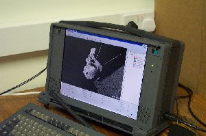
The medical images are segmented using IBM TOSCA (Tool for Segmentation,
Correlation, and Analysys)
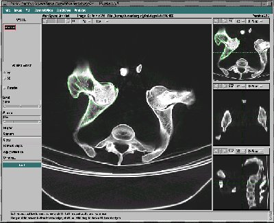
-
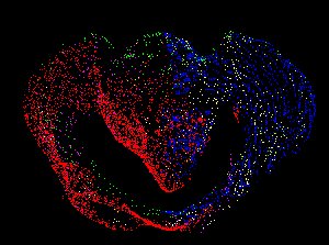
Stack of Contours from TOSCA
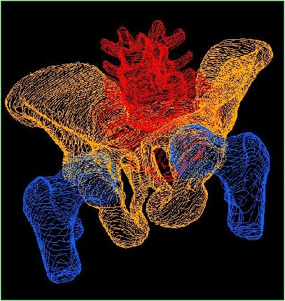
-
Triangulate using INRIA's NUAGES software. Produces VRML v1 file format.
-
Convert VRML v1 into VRML v2.
-
Apply polygon reduction
Method 2


